My dad was my high school physics teacher, so growing up I really liked mathematics and physics. But when I left high school, it was hard for me to decide between physics and fine art. I tried hard to find a combined degree, and I really wanted to do a degree in painting, but somehow, I ended up in a science program at the Australian National University in Canberra.
By the end of my bachelor’s degree, I had had enough of pure science, and so I enrolled in a Master’s degree in painting conservation at the University of Melbourne. I thought it would be a good blend between science and art.
This experience led to a PhD using fluorescence spectroscopy to study the pigments employed throughout the history of art. This type of analysis allowed us to see how a painting was made and aid in the authentication of artwork.
Near the end of my PhD, I went to a workshop at the University of New England in Armidale (NSW) on biological applications of fluorescence, just because it was centred around fluorescence spectroscopy. Professor Enrico Gratton, a world leader in biophysics, was lecturing and he presented on how you could do dynamic measurements of fluorescence in a living cell.
Live cell DNA architecture in real time
I liked how everything became much more complicated by the fact that the sample was alive. I thought that made things more interesting and so I requested a postdoctoral research position (postdoc) with him. Initially, he said, “no, because you’ve never used a microscope”, but added that if I was really keen, I could join his lab as a junior specialist. He gave me a little research project as a test, and since it went well, and I got a paper quite quickly out of that, he let me become a postdoc.
After my postdoc I came back to Australia, first to UNSW in 2013 and then in 2017 I moved to the University of Melbourne.
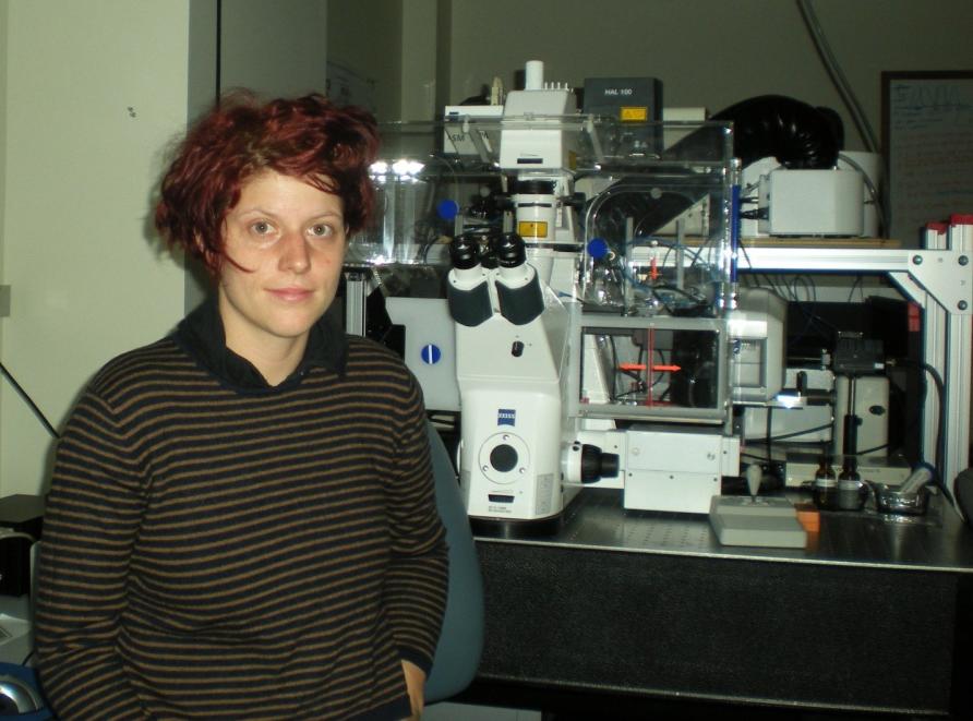
In our lab in the School of Physics, we have a confocal microscope coupled to hardware capable of fluorescence lifetime imaging and an orbital tracking unit. Using this system, we can look at protein-protein interactions on moving objects inside living cells, such as endosomes, DNA double-strand breaks, or the nuclear pore complex.
We also just got a light sheet microscope with single molecule sensitivity that will allow us to look at fast-time intracellular protein trafficking events throughout the cell.
The microscopy methods that we are developing are helping us to better understand how intracellular architecture is organised and the impact it has on protein transport.
This enables cell biologists and medical researchers to interrogate different types of protein dynamics that were previously invisible. These dynamics could include how DNA repair factors arrive at a DNA double-strand break to protect genome integrity and even how viruses hijack intracellular pathways to promote their replication.
In the long term, I hope that we can uncover fundamental insights into how the spatiotemporal organisation of the cell nucleus regulates genome function and DNA target search. This knowledge may be important in processes like stem cell differentiation and cancer development.
Exploring the air we breathe
The data that we acquire looks like noise and is very pixelated, so we’d never win an art competition. But I do love making figures for manuscripts. My sister makes prints for fashion designers so we were thinking it would be cool to use some of the microscopy images or data outputs we generate as a print for clothing.
In 2022 I won the Women in Science Emerging Researcher (WISER) award for Mathematical and Physical Sciences, allowing me to advocate and promote biophysics in Australia. I sometimes lose track of the long-term significance of why I am exploring the development of a particular method of analysis or phenomenon in the cell. And sometimes you can go down obscure pathways, particularly in fundamental research.
I like outreach because people who are not in science always ask you why you’re doing certain things and you’re forced to reflect on the big picture of why you’re doing the research that you’re doing
I think it’s most important for young scientists to be resilient. Everybody has been rejected at multiple points throughout their career, but this is not so obvious from the outside. For example, just prior to the fluorescence spectroscopy workshop in Armidale I had been shortlisted for a conservation scientist position at the National Gallery of Art in Washington, but in the end, I didn’t get it and I was very disappointed.
But then I met Enrico, and with the support of my partner (now husband), I had the chance to do a postdoc with him in California, which was one of the best experiences of my life. So, I think being open-minded to opportunities as they present themselves and not giving up is key.
A lot of students are worried about their career trajectory, but I think you shouldn’t be afraid to change your field a little and take an indirect path. It’s more important to remain interested in your field than to continue begrudgingly.
And maybe you will have to do a bit of extra work to retrain in a new field, but even then, it’s still worthwhile. Skills you have developed in your previous field will always cross over.
– As told to James Carroll and Jeongoh Park, Science Masters students at the University of Melbourne whose studies include Science Communication.
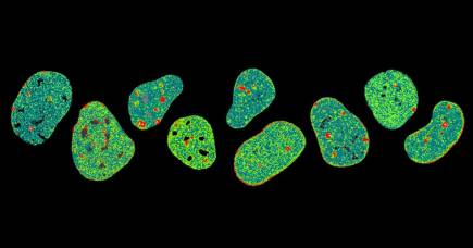
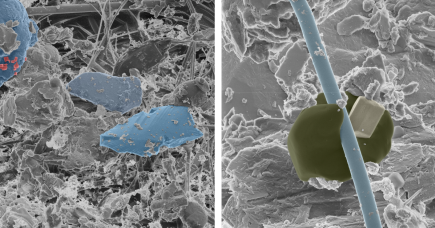
Banner: Fluorescence imaging of live cells. Picture: Supplied